小儿脑干海绵体畸形的处理:在儿童医院工作20多年的经验(Management of pediatric brainstem cavernous malformations: experience over 20 years at the Hospital for Sick Children)
英文摘要:
Object. Because of their location and biological behavior, brainstem cavernous malformations (海绵状血管瘤s) pose a formidable clinical challenge to the neurosurgeon. The optimal management of these lesions requires considerable neurosurgical judgment. Accordingly, the authors reviewed their experience with the management of pediatric brainstem 海绵状血管瘤s at the Hospital for Sick Children. Methods. The authors performed a retrospective chart review of pediatric patients who had received diagnoses of a brainstem 海绵状血管瘤 at the Hospital for Sick Children over the past 20 years. Results. Twenty patients were diagnosed with brainstem 海绵状血管瘤s. The mean age at diagnosis was 10.1 ± 5.4 years, and the patients included 13 boys and 7 girls. The mean maximal diameter of the 海绵状血管瘤 was 14.3 ± 11.2 mm. The lesions were evenly distributed on the right and left sides of the brainstem with 4 midbrain, 13 pontine, and 3 medullary lesions. Seven patients underwent surgery for the management of their 海绵状血管瘤s, with a mean age at presentation of 5.2 years, and a mean 海绵状血管瘤 size of 21.0 mm. Of note from the surgical group, 2 patients had a family history of 海绵状血管瘤s, 2 lesions were medullary, the 海绵状血管瘤 reached a pial surface in 6 of 7 patients, and 6 of 7 lesions were located on the right side. The mean age at presentation among the 13 patients in the nonsurgical group was 12.7 years, and the mean 海绵状血管瘤 size was 10.6 mm. Seven of these patients had a prior history of radiation for tumor, and only 3 had lesions that reached a pial surface. Conclusions. The management of brainstem 海绵状血管瘤s in children is influenced by multiple factors. The majority of patients received conservative management and tended to be asymptomatic with smaller lesions. Patients with larger lesions and direct pial contact, in whom symptoms arose at a younger age were more likely to undergo surgical management. A history of familial 海绵状血管瘤 was also a predictor for receiving surgical treatment. No patients with a prior history of radiation therapy underwent surgery for 海绵状血管瘤s. The presence of multiple lesions seemed to have no impact on the type of management chosen. Patients who underwent surgery did suffer morbidity related to the procedure, and tended to improve clinically over time. Conservative management was associated with new deficits arising in children, some of which improved with time. Consideration of many clinical and radiological parameters is thus prudent when managing the care of children with brainstem 海绵状血管瘤s.
中文摘要:
对象。由于它们的位置和生物学行为,脑干海绵状畸形(海绵状血管瘤s)给神经外科医生带来了较大的临床挑战。这些病变的更佳处理需要相当多的神经外科判断。因此,作者回顾了他们在医院治疗患儿脑干海绵状血管瘤的经验。
方法。作者对过去20年在医院接受诊断的患儿进行了回顾性的图表回顾。
结果。20例患者被诊断为脑干海绵状血管瘤。平均年龄(10.1±5.4)岁,其中男生13例,女生7例。脑干海绵状血管瘤的平均较大直径为14.3±11.2 mm。病变均匀分布于脑干左右,中脑4例,桥13例,髓质3例。7例患者接受了脑干海绵状血管瘤手术治疗,平均年龄为5.2岁,平均脑干海绵状血管瘤大小为21.0 mm。手术组有2例有脑干海绵状血管瘤家族史,2例为髓样病变,7例中有6例脑干海绵状血管瘤达软脑膜面,7例中有6例位于右侧。非手术组13例患者的平均发病年龄为12.7岁,平均脑干海绵状血管瘤大小为10.6 mm。这些患者中有7人有肿瘤放射史,只有3人的病灶达到了软脑膜表面。
结论。儿童脑干海绵状血管瘤的管理受到多种因素的影响。大多数患者接受保守治疗,往往无症状,病变较小。有较大病变和直接颈部接触的患者,其症状在较年轻时出现,更有可能接受手术治疗。家族性脑干海绵状血管瘤史也是接受手术治疗的评估因素。既往有放疗史的患者均未行脑干海绵状血管瘤手术。多发性病变的存在似乎对所选择的治疗类型没有影响。接受手术的患者确实会出现与手术相关的发病率,而且随着时间的推移,其临床症状往往会有所好转。保守治疗与儿童新出现的缺陷有关,其中一些随时间而好转。因此,在管理脑干海绵状血管瘤患儿的护理时,应谨慎考虑许多临床和放射学参数。(DOI: 10.3171 / 2009.6.peds0923)
海绵状血管瘤是比较常见的脑部病变,约占总人口的0.5%,占全部脑血管畸形的5-10%。大体病理分析,这些病变是清晰的分叶状肿块,呈桑树状。它们由薄壁的血管通道组成,呈蜂窝状,通常由一个胶质细胞平面从正常大脑中分离出来。先前研究的作者已经确定了2种主要的海绵状血管瘤:一种以单个病变为特征的散发型,一种以多发性病变为表现的常染色体显性家族型。在家族性海绵状血管瘤中,病变被观察到是新生的。这与之前认为海绵状血管瘤只是先天性病变的观点相反。
尽管海绵状血管瘤在组织学上是良性的,但由于出血和扩张引起的肿块效应,它能导致毁灭性的神经损伤。特别是位于脑干的海绵状血管瘤患者,由于基本脑干核和上行/下行束的中断,存在较高的死亡和残疾概率。脑干海绵状血管瘤并不少见,因为据报道脑干海绵状血管瘤占全部脑干海绵状血管瘤的35%。一例报道的脑干海绵状血管瘤切除术是80多年前由Walter Dandy实施的。从那时起,微神经外科技术取得了较大的进步,脑干手术方法也有了很大的创新。随着这些进展,管理这些复杂病变的决策算法也在不断发展。脑干海绵状血管瘤在儿童时期表现出独特的临床困境。一方面,脑干海绵状血管瘤的GTR提供了一个完全治愈和解决未来灾难性事件风险的机会。这种益处在儿童中可能被放大,因为较长的寿命意味着更大的潜在出血风险。然而,也需考虑死亡的直接风险和手术并发症。考虑到这些因素,我们被要求回顾我们在医院治疗患儿脑干海绵状血管瘤的经验。
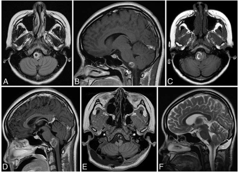
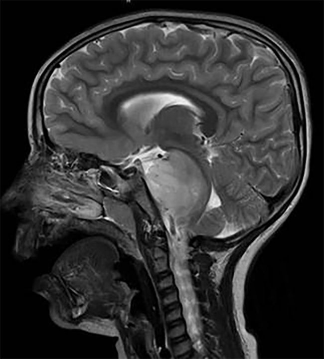
图示:一次出血(A和B)后获得轴位和矢状位t1加权FLAIR MR图像,3周后再次获得(C和D),临床恶化。病变在间隔时间内直径增加了10毫米。轴向t1加权FLAIR MR图像(E)和矢状t2加权MR图像(E)也显示在GTR后数月。
我们回顾了过去20年加拿大多伦多儿童医院对患病儿童脑干海绵状血管瘤的管理。桥脑海绵状血管瘤是较常见的。三分之二的患者接受保守治疗,其中一半患者无症状。全部接受手术的病人都有病变的症状。在我们的经验中,较大的脑干海绵状血管瘤与软膜瘤接触,在较年轻、有症状的患者中被诊断,更有可能被手术治疗。先前的放射治疗和海绵状血管瘤多样性均未影响手术的处理。术后发病率可能是的,但在术后的时间内,通常有的临床好转机会。因为一个患有脑干海绵状血管瘤的孩子有可能活上几十年,所以需仔细权衡未来潜在的出血事件和手术并发症的风险。
该研究主要成员之一为INC国际神经外科医生集团旗下国际神经外科顾问团(WANG)成员James T. Rutka教授,担任国际神经外科学院院长(2011-2014),多伦多大学儿童病院、亚瑟和索尼亚拉巴特脑瘤研究中心主任(1998年至今)。James T. Rutka教授曾与名古屋大学的杉田健一郎(Kenichiro Sugita)博士在微血管神经外科领域开展了临床研究,并于1990年在东京骏天大学(Juntendo University)获得了分子免疫学博士后研究奖学金。教授发表了超过500多篇的文章,在临床上的研究方向以颅内肿瘤为主,对胶质瘤、纤维瘤、颅咽管瘤、室管膜瘤具有多年的临床经验,擅长清醒开颅术及显微手术。在较近的临床研究上,教授又将重点集中在了儿童脑瘤和癫痫的外科治疗上,并取得了合适的成果。
- 文章标题:小儿脑干海绵体畸形的处理:在儿童医院工作20多年的经验
- 更新时间:2019-11-27 17:15:47

 400-029-0925
