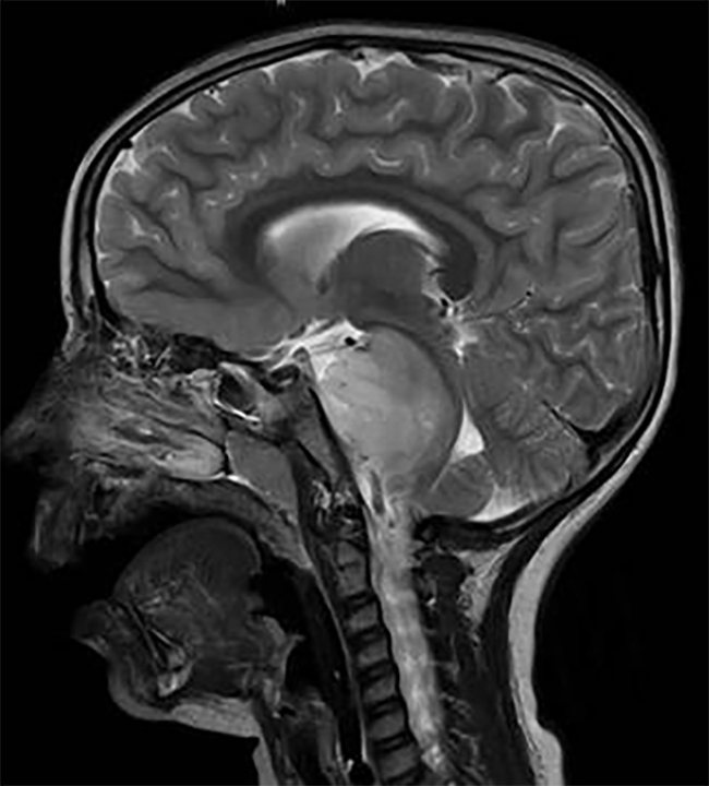
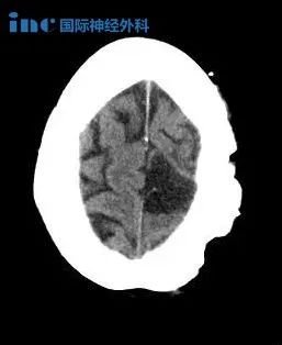
颅内原发性神经节胶质瘤(Pediatric intracranial primary anaplastic ganglioglioma)
英文简介:
Background Primary intracranial anaplastic gangliogliomas are rare tumors in the pediatric patient group. Most of them present with symptoms of elevated pressure or symptomatic epilepsy. Extraaxial location is far more common than axial location. On MRI examination, they mimic pilocytic astrocytomas. The outcome after surgery depends mainly on the possible amount of surgical resection, and oncological therapy is necessary to prevent recurrence of the disease. Case report An 11-year-old boy presented with headache and double vision due to obstructive hydrocephalus. MRI of the brain revealed an axial partially contrast enhancing lesion in the quadrigeminal plate extending from the cerebellum to the pineal gland and causing hydrocephalus. Subtotal removal of the lesion was performed, and the diagnosis of an anaplastic ganglioglioma was established and confirmed by the reference center. At the latest follow up (3 months), the boy is without any neurological symptoms and scheduled for radiation therapy as well as chemotherapy
中文简介:
背景:原发性颅内间变性神经节胶质瘤在儿科患者中是少见的肿瘤。大多数患者表现为血压升高或癫痫症状。轴外定位远比轴向定位常见。在核磁共振检查,他们模拟毛细胞星形细胞瘤。手术后的结果主要取决于可能的手术切除量,肿瘤治疗是必要的,以防止疾病的复发。
病例报告1例11岁男童因梗阻性脑积水而出现头痛及复视。大脑核磁共振成像显示,轴向部分对比增强病变在神经板从小脑延伸到松果体,并导致脑积水。对病变进行次全切除,诊断为间变性神经节胶质瘤,并由参考中心予以确认。在较近的跟进(3个月),男孩没有任何神经系统症状,并计划接受放射治疗和化疗。
神经节胶质瘤是一种中枢神经系统的肿瘤,怀疑起源于错构瘤性神经胶质神经病变的常见前体细胞。组织病理学上,这些肿瘤由神经节细胞和胶质细胞组成。它们在全部小儿中枢神经系统肿瘤中占4%,因此很少见。间变性颅内神经节胶质瘤是一种侵袭性神经节胶质瘤亚型,主要表现为有丝分裂、纤维增生、血管增生和坏死。BRAF V600E突变在颅内间变性神经节胶质瘤中比在脊髓髓内病变中更常见。报告原发性良性神经节神经胶质瘤的继发性恶变。
神经节神经胶质瘤在1926年一次被描述为一种独特的颅内肿瘤。2011年报道的规模较大的一系列间变性神经节胶质瘤有85例,中位年龄为25.5岁,较常见的时间位置(27%),中位总生存期为28.5个月。
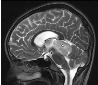
图示:术前矢状面t2加权像显示神经板高强度病变(星形),从小脑延伸至丘脑。
INC国际神经外科医生集团旗下组织国际神经外科顾问团(WANG)成员巴特朗菲教授作为主要研究者之一,擅长大脑半球病变、脑干病变、脑血管疾病、脑内深层区胶质瘤、颅颈交界处的病变等的肿瘤切除术、神经吻合术以及各种椎管内肿瘤切除术,以高超的技术手法和顺利前提下高切除率手术而,在中国患者群中被尊称为“巴教授”。
- 文章标题:颅内原发性神经节胶质瘤
- 更新时间:2019-11-06 17:59:15

 400-029-0925